

 News Center
1.Background
In early 2025, high-parameter flow cytometers and other biotechnology equipment were added to export licensing control lists. Following the tightening of import channels, research institutions dependent on imported systems have generally faced uncertainty in equipment maintenance and reagent supply, with the continuity of clinical sample testing and data security coming under direct impact. Establishing autonomous and controllable tissue imaging capabilities has shifted from a long-term plan to an urgent necessity.
At the same time, tissue pathology research itself is confronting a generational bottleneck. The challenge of obtaining richer molecular information from a single tissue section while preserving the original spatial structure—a quest spanning three generations of technology—remains only partially resolved.
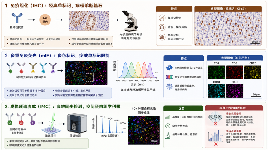
First-generation immunohistochemistry established the cornerstone of single-marker detection
Through specific antibodies and DAB chromogenic staining, pathologists can directly assess the presence and intensity of protein expression under a microscope—still a core tool for tumor subtyping and companion diagnostics. However, it is inherently single-marker detection: one section answers only one protein question, and serial sectioning strategies cannot guarantee precise cell-to-cell positional correspondence across different sections.
Second-generation multiplex immunofluorescence overcomes the limitations of single-labeling
Different fluorescent dyes enable simultaneous detection of three to five proteins on a single section. However, when the parameter count exceeds five, signal crosstalk increases sharply, and the number of protein markers that can be stably detected in practice struggles to exceed single digits.
Third-generation mainstream imaging mass cytometry (IMC) replaces fluorescent dyes with metal-tagged antibodies
By relying on mass spectrometry detection, it overcomes the constraints of spectral overlap, enabling high-dimensional simultaneous detection of over forty protein markers on a single section. Nevertheless, its methodological framework confines detection targets to the mass range of exogenous tag elements, leaving endogenous metals outside routine workflows without standard detection protocols. The system is closed, preventing users from deeply adjusting sampling parameters and detection settings. Quantitatively, it outputs only relative signal intensity without support for standard introduction, making data incomparable across batches and laboratories.
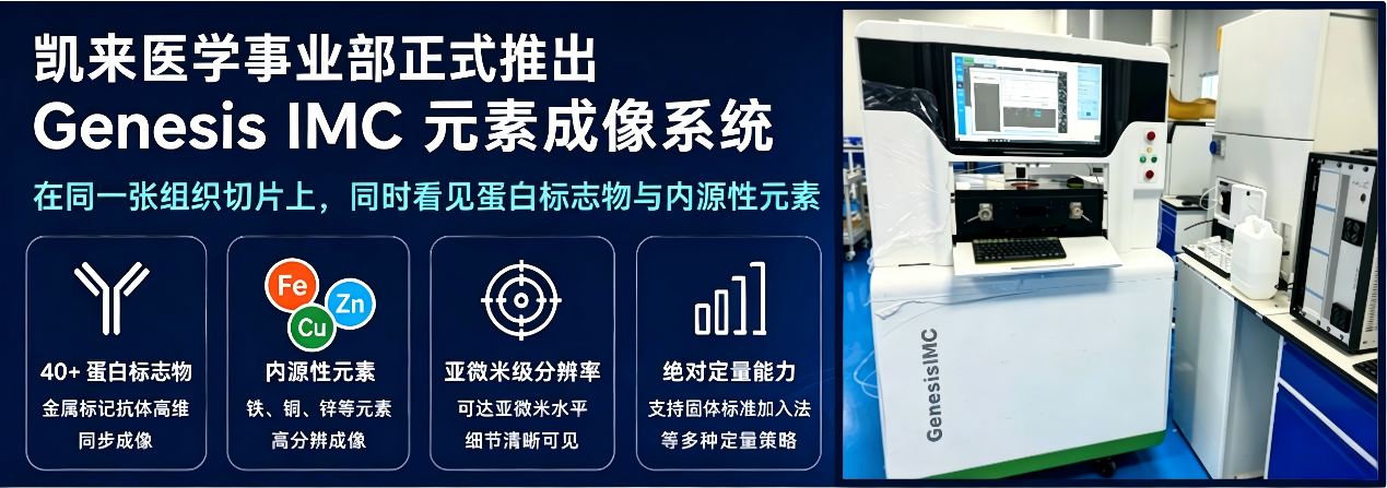
The convergence of external supply chain risks and internal technical ceilings demands a new pathway that combines autonomous control with methodological extensibility. On this basis, Chemlab Medical Business Unit officially launches the Genesis IMC Elemental Imaging System, providing full-chain support from instrument hardware to testing services for research applications. With a dual-drive positioning of instruments plus services, we are committed to continuous hardware upgrades, software ecosystem enrichment, and expansion into clinical application scenarios.
2.System Composition
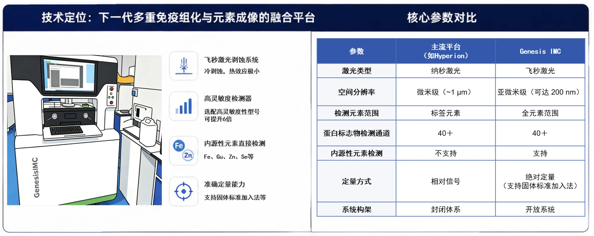
The Genesis IMC system consists of the following three core modules.
Hardware Module: Features a femtosecond laser sampling system coupled with a high-sensitivity mass spectrometer detector. The cold ablation effect of the femtosecond laser minimizes the heat-affected zone, which is critical for preserving fine biological tissue structures.
Service Module: Fully open metal imaging testing services supporting sample submission. Additionally, we offer open hardware parameter and experimental condition optimization services, supporting collaborative teams to jointly optimize labeling workflows, establish endogenous element calibration methods, and develop multimodal data fusion algorithms.
Data Module: Provides full-process data output from raw mass spectrometry data to spatial imaging maps, supporting custom algorithm development and in-depth analysis by users based on open data formats.
3.Core Highlights
The following four dimensions illustrate the breakthroughs of Genesis IMC over previous-generation technologies.
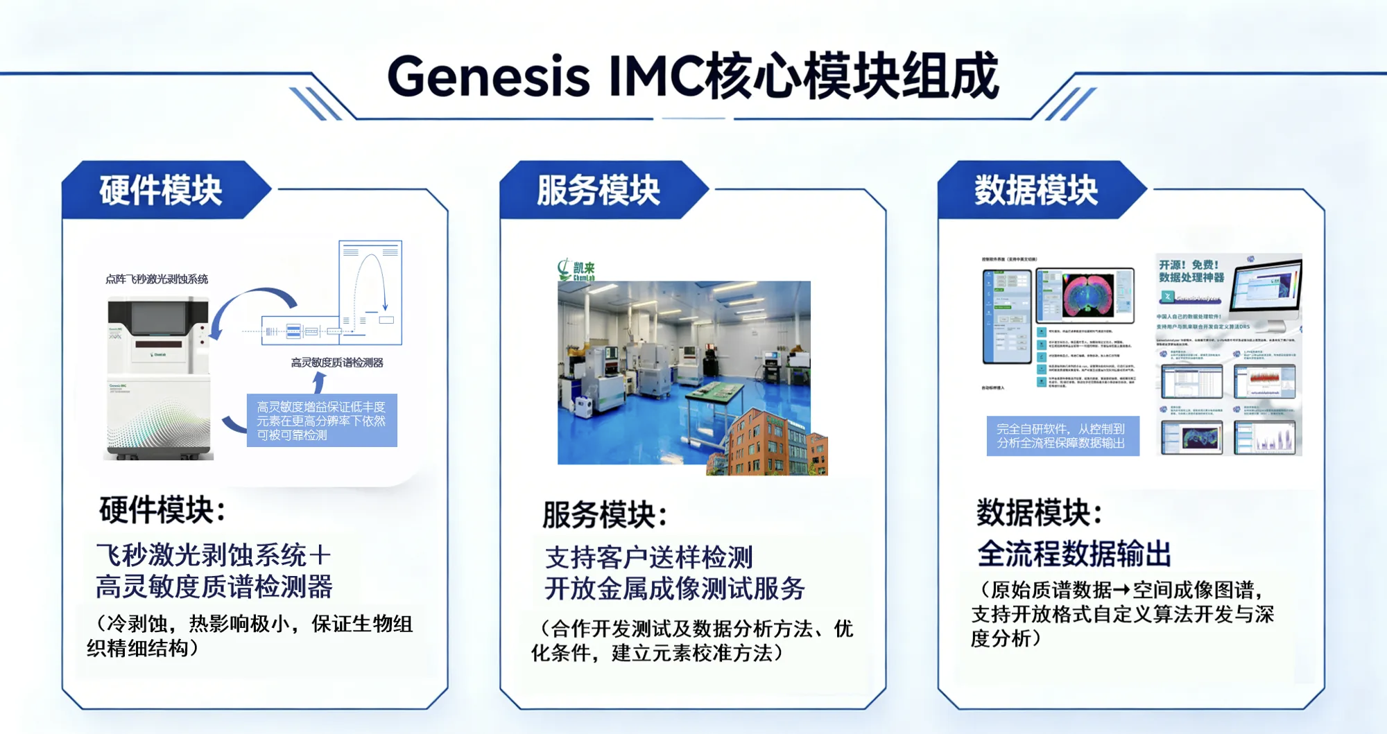
Highlight 1: Endogenous elements – no labeling required, directly visible
Traditional immunohistochemistry and multiplex immunofluorescence rely entirely on exogenous labels and cannot capture tissue-intrinsic elemental information. Although mainstream IMC platforms enable metal-tag detection, their mass windows and calibration systems are specifically optimized for tag elements. Tissue-endogenous elements such as iron, copper, and zinc lack standard detection protocols, and endogenous metal metabolic signals are not incorporated into routine analytical workflows.
Genesis IMC does not require commercial antibody kits; it directly detects endogenous elements inherent to tissues. Preliminary comparative tests demonstrate that protein marker imaging quality is comparable to mainstream platforms, while simultaneously achieving high-resolution imaging of endogenous elements including iron, copper, and zinc. Researchers can now read both the spatial expression profiles of protein markers and the elemental metabolic microenvironment from the same tissue section.
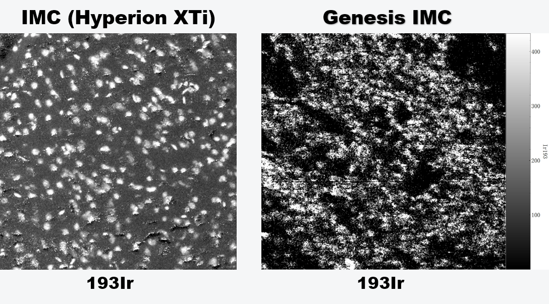
Note: IMC imaging data in this document were provided by client institutions and are used solely for this technical comparison demonstration. They may not be repurposed or republished without permission. Elements not presented in Genesis IMC images were not included in this comparison and do not imply the system's inability to detect them.
Parallel testing on the same tissue across two platforms shows that Genesis IMC achieves high consistency with mainstream platforms at the protein marker level. Each rare-earth metal-tagged antibody channel displays clear tissue distribution contours, with comparable signal contrast and spatial localization capabilities, demonstrating imaging resolution on par with mainstream platforms.
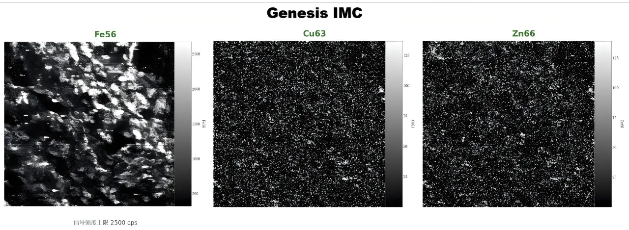
Genesis IMC Breaks Through the Quality Limits of Label Elements on Mainstream Platforms,with a detection range unconstrained by tag elements. In this test, endogenous elements including iron, copper, and zinc were clearly resolved: iron delineates tissue morphology, while copper and zinc distributions exhibit complementary spatial patterns with protein markers, providing an independent dimension for microenvironment interpretation.
Highlight 2: Spatial resolution extended from micrometer to sub-micrometer levels
Immunohistochemistry and multiplex immunofluorescence are limited by the optical diffraction limit, typically achieving micrometer-level resolution. Mainstream IMC platforms operate at approximately 1 micrometer resolution, which is the conventional upper limit of their hardware architecture.
Genesis IMC delivers comparable imaging quality at the same resolution. However, micrometer resolution is not the endpoint for Genesis IMC. Under optimized experimental conditions, the platform achieves sub-micrometer spot sizes, enabling further refinement in subcellular elemental localization. With higher-sensitivity detector models, low-abundance elements remain reliably detectable even at higher resolutions.
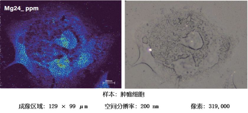
Benchmark tests were conducted at micrometer resolution, but the upper limit of Genesis IMC extends beyond this. The platform has already achieved subcellular Mg24 imaging at 200 nm spot size, with further localization precision possible.
Highlight 3: Traceable absolute concentration quantification
In clinical pathology research and biomarker validation, quantitative reliability directly determines the credibility of research conclusions. Immunohistochemistry has long relied on semi-quantitative scoring systems, such as H-scores or positive percentage. Mainstream IMC platforms output relative signal intensity—semi-quantitative data—without support for standard introduction. Detection signals are subject to multiple interferences including tissue density, section thickness, and laser energy fluctuations, making it difficult to establish a unified comparison baseline across different batches and laboratories. For biomarker studies requiring multi-center validation, this quantitative incomparability poses a substantial obstacle.
Genesis IMC supports the solid standard addition method, in which multi-element standards of known concentration are deposited on the tissue surface to establish calibration curves, converting signal intensity to absolute concentration under matrix-matched conditions. It also supports matrix-matched standard methods and internal standard dynamic correction, directly reflecting the metabolic status and functional changes of endogenous metal elements.
The gel standard method is a distinctive feature of the Genesis IMC quantification system: gelatin or agarose is used as the matrix, liquefied, spiked with metal standard solutions, cooled and sectioned to produce standard samples with controllable thickness and homogeneous elemental distribution. Its advantages include: avoiding ablation variability caused by biological tissue heterogeneity; clean background with trace element interference further reduced via metal-binding resins; and mold-based molding ensuring preparation reproducibility. This open quantitative strategy enables cross-laboratory comparability for absolute concentration measurements of endogenous metal elements.
For protein markers, although their absolute quantification pathway via indirect metal-tag detection differs from that of endogenous elements, the platform's open architecture permits the introduction of exogenous standards on the section surface or in co-deposited layers, offering the potential to establish traceable quantitative frameworks for protein expression.
Highlight 4: From standardized protocols to an open collaboration system architecture
Mainstream IMC platforms have fully solidified workflows, leaving users with limited ability to deeply customize front-end labeling strategies or back-end algorithms. When research extends toward metal metabolism, elemental speciation analysis, or endogenous ion regulation, it becomes difficult to make deep adjustments in detection range or methodological parameters.
Genesis IMC allows adjustment of sampling parameters, optimization of gas flow conditions, development of labeling protocols, and even reconstruction of data analysis pipelines to address specific scientific questions.
4.Open Collaboration
The Genesis IMC Elemental Imaging System is now officially launched, and our metal imaging testing services are fully open. We sincerely invite collaboration with the following partners:
1.Research teams with biological samples and data analysis capabilities
In fields such as tumor immune microenvironment, neurodegenerative diseases, or developmental biology, where protein marker imaging data comparable to mainstream platforms is required along with exploration of endogenous metal element distribution patterns—sample submission testing is welcome.
2.Teams engaged in front-end labeling strategy or back-end imaging algorithm research
Chemlab is willing to open hardware parameters and experimental conditions to jointly optimize labeling workflows, establish endogenous element calibration methods, and develop multimodal data fusion algorithms.

Conclusion
Chemlab Genesis IMC is committed to providing a more extensible technological pathway for the metal imaging field. As iron, copper, and zinc in tissues are visualized with the same precision as protein markers for the first time, new possibilities are opening at the intersection of metallomics and spatial proteomics.
Chemlab looks forward to working with all researchers to advance the implementation and evolution of this methodology. For testing inquiries and collaboration discussions, please feel free to contact us at any time.
Copyright 2024 Shanghai CHEMLAB Instrument Co., Ltd. All Rights Reserved
Shanghai ICP Filing No.65656565