

 News Center
Research Background
Otoliths are calcified structures in teleost fishes, formed during the embryonic stage. They are acellular and metabolically inert, yet exhibit high interspecific morphological variability. As continuously growing biomineralized structures throughout the fish's life, otoliths record environmental information experienced during individual growth with high temporal resolution. Their trace element composition and isotopic signatures have been widely applied in studies of fish age and growth determination, early life history reconstruction, migratory route analysis, and habitat use. In particular, elemental ratios such as Sr/Ca, Ba/Ca, and Mg/Ca are commonly used as indicators of different water mass structures, salinity gradients, and depth changes, serving as crucial links between fish ecological processes and marine environmental changes.
With the advancement of fish ecology and fisheries resource management research towards greater precision and process-orientation, traditional otolith trace element chemistry analysis techniques have gradually revealed limitations in spatial resolution, temporal resolution, and quantitative accuracy. Although Laser Ablation-Inductively Coupled Plasma-Mass Spectrometry (LA-ICP-MS) has become a routine method for otolith microchemical analysis, conventional nanosecond lasers, such as 266 nm, 213 nm, and 193 nm systems, exhibit significant thermal and matrix effects. During ablation, they are prone to causing rim melting, elemental migration, and quantitative deviations. These limitations are particularly pronounced when analyzing fine growth increments during early life stages at high spatial resolution for imaging and precise quantification, significantly compromising data reliability.
The Game-Changing Solution
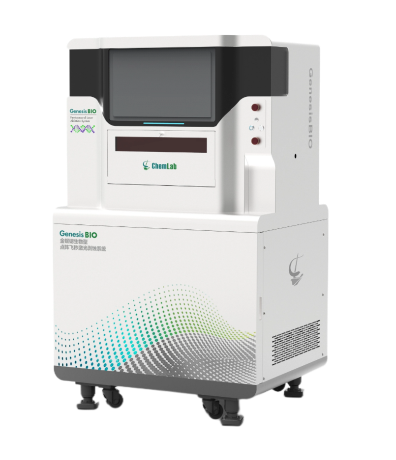
GenesisBIO Matrix-Array Femtosecond Laser Ablation System
The GenesisBIO Matrix-Array femtosecond laser ablation system, fully In-house Development and manufactured by Chemlab Instrument Co., Ltd. (Shanghai), coupled with mass spectrometry, offers significant advantages over nanosecond laser-MS systems. It enables more detailed resolution of elemental variation patterns from the core to the edge of fish otoliths, allowing finer characterization of an individual's environmental history from hatching, through larval and juvenile stages, to adulthood. Key advantages include:
Ultra-Low Thermal Effects, Enabling "Cold Ablation"
With a pulse width of <260 fs, the pulse duration is on the order of 10-15seconds. The ablation process is a "Coulomb explosion" cold ablation mechanism. The energy deposition occurs faster than thermal diffusion, effectively suppressing rim melting around the ablation pit on the otolith. By inhibiting the thermal decomposition of the calcium carbonate matrix, it effectively suppresses the migration of microchemical elements within the otolith, enabling high-spatial-resolution ablation and precise analysis of microchemical element changes during critical early life history stages.
<1 μm High-Resolution Observation of Otolith Increments
An integrated coaxial high-definition color observation system with an optical resolution of ≤1 μm and high-brightness transmitted light illumination significantly enhances the observation and reading of otolith cross-section increments, effectively improving the accuracy of age determination for larval and juvenile fish otoliths.
Breaking the Spatial Resolution Limit
Possessing an ultra-high spatial resolution ablation capability of ≤1 μm, enabling sub-micrometer-scale precise analysis. It breaks through the limitations of traditional laser ablation in identifying fine structures, allowing for more detailed and higher-resolution analysis of otolith microstructure and elemental distribution. This provides more reliable data support for studies on fish life history reconstruction and environmental response.
Analytical Highlights
1. Sample Collection and Preparation
Target fish samples are selected, and their sagittal otoliths are extracted. After removal, otoliths are repeatedly rinsed with ultrapure water to remove adhering soft tissues and minimize surface contamination risks. Cleaned otoliths are air-dried naturally and set aside. The otoliths are then embedded in epoxy resin, sectioned along the core-to-edge growth axis, and progressively polished until a flat surface with clearly visible structures is obtained. All samples are stored in a clean environment prior to analysis.
2. Line Scan Trend Analysis
The Shanghai Chemlab Spectrum Laboratory employed the fully Independent Research and Development GenesisBIO Matrix-Array femtosecond laser ablation system coupled with a triple quadrupole ICP-MS to perform line scan analysis on the otoliths of Antarctic fish from the core to the edge. This successfully yielded continuous, stable multi-element signals and elemental ratio sequences.
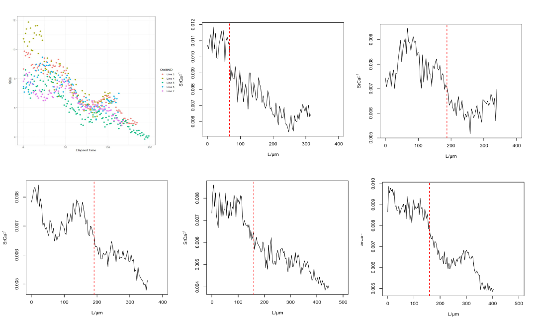
Sr/Ca variation trend from otolith core to edge
Regarding result stability, the GenesisBIO system exhibited no significant signal spikes or anomalous peaks throughout the entire line scan process. The Sr/Ca ratio variations corresponded well with the otolith growth structures. This demonstrates that femtosecond laser ablation effectively reduces the elemental fractionation commonly observed in traditional nanosecond laser systems, particularly minimizing the selective volatilization of light versus heavy elements caused by localized high temperatures, thereby enhancing the authenticity and comparability of otolith microchemical data.
During the analysis, due to the extremely short pulse duration of the GenesisBIO laser, energy deposition in the material is predominantly a non-thermal mechanism. This effectively suppresses melting, recrystallization, and thermal diffusion effects within the otolith calcium carbonate matrix during ablation. The line scan path boundaries are clear, and the ablation width and depth are highly controllable, ensuring good temporal continuity and spatial consistency of elemental signals along the growth axis. This is particularly critical for otolith line scan analysis, which emphasizes "time-series integrity."
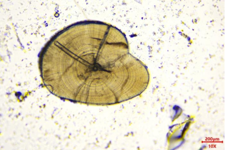
Schematic of otolith cross-section under an optical microscope; the grey line indicates the ablation path.
3. 1 μm High Spatial Resolution Otolith Mapping (2D Imaging) Analysis
For otolith imaging analysis, the GenesisBIO system performs point-by-point ablation, reconstructing elemental signals into two-dimensional distribution images that visually display microchemical differences across different regions of the otolith. The entire otolith cross-section is encompassed within the imaging area, with parameters for spacing, spot size, and single-point ablation time set as per experimental conditions. The system automatically performs sequential point-by-point ablation and multi-element signal acquisition according to a preset grid pattern. In this experiment, the GenesisBIO system was coupled with an inductively coupled plasma time-of-flight mass spectrometer (ICP-TOF-MS), using helium as the carrier gas to measure the microchemical composition of the otolith cross-section.
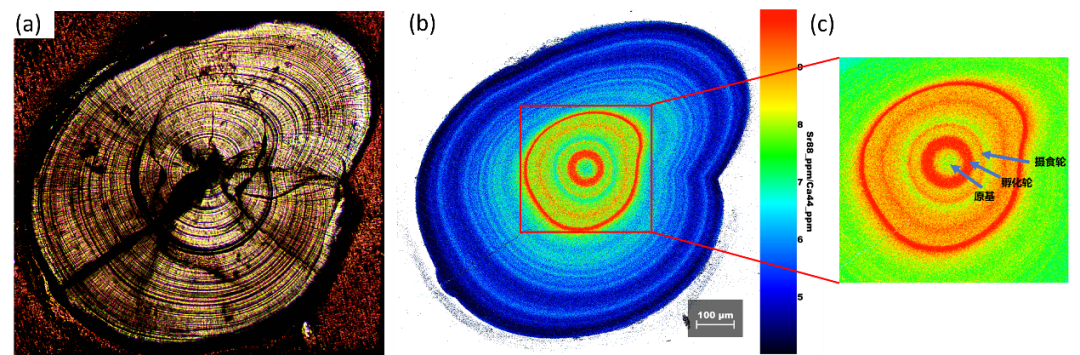
a. Microscopic image of Antarctic fish otolith cross-section; b. Sr/Ca 2D elemental map of the otolith (length: 978 μm, width: 920 μm, spatial resolution: 1 μm, pixels: 899,760); c. Key life history rings in the otolith core.
The results show that the otolith growth ring structures are clear, continuous, and have well-defined boundaries, demonstrating good measurability. During imaging, the spatial structures and microchemical distribution of the otolith core and growth rings exhibited strong correspondence, achieving a "what you see is what you get" imaging effect. The boundaries of the growth rings are distinct and measurable, significantly enhancing the correlation between otolith microstructure and microchemical signals, overcoming the traditional limitation of "decoupling between structure and chemical signals" in otolith microchemical analysis.
Conclusion
Otolith imaging analysis employs point-by-point ablation using the GenesisBIO system, reconstructing elemental signals into 2D distribution images that visually represent microchemical variations across different regions of the otolith. The entire cross-section is included in the imaging area, with spacing, spot size, and single-point ablation time set accordingly. The system performs automated sequential ablation and multi-element signal acquisition based on a preset grid. In this experiment, the GenesisBIO system was coupled with ICP-TOF-MS, using helium as carrier gas to measure the microchemical composition of the otolith cross-section.
Instrument Product Information
GenesisBIO Matrix-Array Femtosecond Laser Ablation System
Headquartered in the Marine Science and Technology Innovation Park, Lingang New Area, Shanghai, Chemlab Instrument Co., Ltd. focuses on the independent R&D and intelligent manufacturing of high-end analytical instruments. Leveraging its core Matrix-Array femtosecond laser ablation technology, Chemlab provides comprehensive precision testing solutions for various sectors, including high-end semiconductor manufacturing, cutting-edge scientific research, and precision medical diagnostics.
Copyright 2024 Shanghai CHEMLAB Instrument Co., Ltd. All Rights Reserved
Shanghai ICP Filing No.65656565