

 News Center
Research Background
In the abyssal sediments of the Clarion–Clipperton Zone (CCZ) in the eastern Pacific, a silicate mineral named phillipsite occurs frequently and exhibits a certain spatial coupling relationship with rare earth element (REE)-enriched layers. This phenomenon led early researchers to regard it as an important carrier mineral of REEs, with some even suggesting that phillipsite is one of the key mineral phases controlling REE enrichment in deep-sea sediments.
However, with the advancement of in situ microanalytical techniques, a large body of measured data has shown that the total REE content of phillipsite itself is generally less than 200 ppm, contrasting sharply with its ubiquitous presence in REE-rich layers—this contradiction has prompted the academic community to re-examine the true role of phillipsite in the REE enrichment process: is it an "enricher" of REEs, or merely an "accompaniment" of specific sedimentary environments?
This study conducts systematic mineral morphological and in situ geochemical analyses on phillipsite separated from a core in the western CCZ of the eastern Pacific, aiming to answer this core scientific question and provide new theoretical support for the exploration and evaluation of deep-sea REE resources.
Research Highlight—Breakthrough Application of High-Spatial-Resolution fsLA-ICP-MS Elemental Mapping Using GenesisGEO
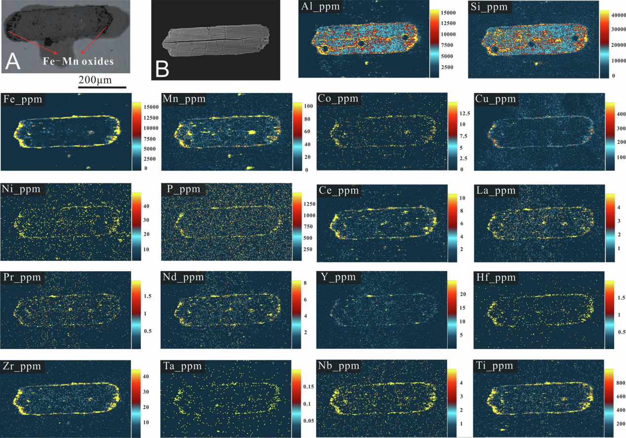
The key technical approach in this study is fsLA-ICP-MS elemental mapping imaging on single phillipsite particles. This technique was performed at Chemlab Pro Technologies using a GenesisGEO system with a 2 μm laser spot size and 100 Hz ablation frequency for surface scanning, coupled with a TOFWERK icpTOF mass spectrometer for simultaneous acquisition of all elements, ultimately generating micron-scale spatial distribution images of elements (Fig. 1). This method visually presents the fine spatial distribution patterns of REEs and related elements within phillipsite particles.
Key Findings from Imaging Results:
(1)Spatial distribution of REEs is highly consistent with that of Fe-Mn oxides
The elemental mapping images clearly show that the enrichment areas of Ce, La, Pr, Nd, Y, and other REEs completely coincide with the spatial distributions of Fe, Mn, Co, Ni, Cu, and other typical indicator elements of Fe–Mn oxides. All are concentrated at the outermost edges of phillipsite particles and in surface microfractures, while signals in the interior core regions are weak or even absent. This "co-localization" feature in space provides the most direct and compelling evidence that iron oxides control REE enrichment—REEs are not distributed within the phillipsite lattice, but are instead attached to the Fe–Mn oxide phases on the particle surfaces.
(2) Iron-dominated, phosphorus-assisted REE contribution model
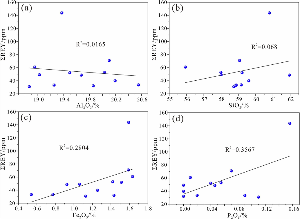
The mapping images also reveal that although P is also mainly distributed at the edges of phillipsite particles, its spatial extent differs somewhat from that of Fe, Mn, and REEs, and its enrichment intensity is significantly lower than that of Fe. Combined with the correlation differences between ΣREY and Fe₂O₃ versus P₂O₅ from spot analyses (Fig. 2), this indicates that iron oxides are the primary source of REEs in phillipsite, with phosphate contributing secondarily.
This conclusion is directly validated at the spatial scale—the high-signal areas of Fe on the imaging maps correspond exactly to the high-signal areas of Ce, and Ce accounts for more than 65% of ΣREY at its highest. This is the typical geochemical behavior of iron oxides that preferentially oxidize and adsorb Ce3+ to form Ce4+enriched phases.
(3)Visualization of the "adsorption channels" within phillipsite internal structure
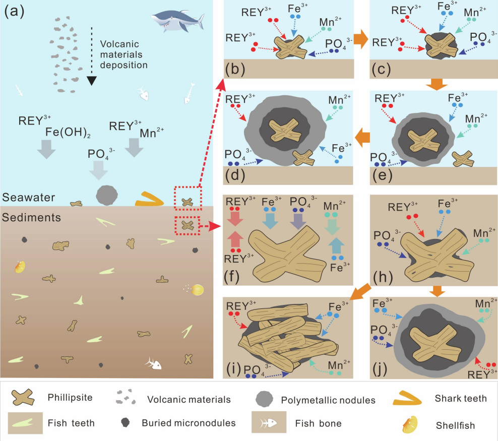
The imaging maps further reveal numerous microfractures and dissolution pores developed on the surfaces of phillipsite particles—these pore structures are precisely the physical channels through which Fe–Mn–P oxides migrate from pore water into the particle interiors. The signal intensities of Fe, Mn, P, and REEs are significantly higher on both sides of the fractures than in areas far from the fractures, intuitively confirming the strong adsorption capacity of the porous phillipsite structure for oxide particulates from the surrounding water (Fig. 3). It is precisely this adsorption process that causes phillipsite, which has extremely low REE content itself (average ΣREY of only 54 ppm), to be coated with a surface layer of iron oxide film with considerable REE content, creating the illusion of "false" enrichment in bulk analyses.
Conclusions
With the aid of high-spatial-resolution fsLA-ICP-MS elemental mapping using the GenesisGEO system, this study visually reveals, at the micron spatial scale, the true occurrence state of REEs in phillipsite particles—they are almost entirely concentrated in the Fe–Mn oxide films on the particle surfaces, rather than being distributed within the phillipsite crystal lattice. This finding fundamentally revises the traditional view that "phillipsite is an important carrier of deep-sea REEs," and clearly indicates that the so-called "REE enrichment" is essentially the result of adsorption and incorporation of iron oxides, rather than an inherent mineralogical property of phillipsite itself.
Paper Information
Zhongrong Qiu,Chunhui Tao, Yinan Deng, et al, Geochemistry of phillipsite in abyssal sediments from eastern pacific: indications of false rare earth element enrichment, Ore Geology Reviews,Volume195,2026,107388,ISSN 0169-1368,https://doi.org/10.1016/j.oregeorev.2026.107388.
Copyright 2024 Shanghai CHEMLAB Instrument Co., Ltd. All Rights Reserved
Shanghai ICP Filing No.65656565